Friday, April 25, 2014

Malignancies

Malignancies-

-Acute/Chronic Lymphocytic Leukemia-


-Acute Leukemia is the most common form of cancer in children (30 percent of all children malignancies)

-Acute Lymphocytic Leukemia (ALL) is 5 times more common than AML

-The overall survival rate is 85% with ALL

-The most common presenting symptoms of ALL are fever, bleeding, bone pain, and lymphadenopathy.

-Most children with ALL with have anemia and/or thrombocytopenia with normal or depressed WBC

-Bone marrow biopsy is indicated if atypical cells are found in peripheral blood, use explained depression of more than one peripheral blood element, or unexplained lymphadenopathy or hepatosplenomegaly with associated cytopenias

-The diagnosis of CNS leukemia requires confirmation of leukemic cells in the CSF

-Treatment protocols are multi drug and divided into phases of induction, consolidation, and maintenance
-Most treatment protocols take 2-3 years to complete
-There is therapy also directed at the central nervous system


-Chronic Lymphocytic Leukemia (CLL)-
-B Cell Chronic Lymphocytic Leukemia is considered to be identical to the mature B cell neoplasm small lymphocytic leukemia

-CLL is characterized by a accumulation of functionally incompetent lymphocytes which are monoclonal in origin

-Most patients present with painless generalized lymphadenopathy which has been present usually for years

-Hepatosplenomegaly and extranodal infiltrates may be seen

-Patients on the peripheral blood smear with normal or mild lymphocytosis but will have an absolute lymphocyte count of less than 5000/microL

-A serum paraprotein or hypogammaglobulinemia is found in 20-40 percent of the cases

-Patients with CLL patients may develop autoimmune hemolytic anemia, pure red cell aplasia, and autoimmune thrombocytopenia

-Elevations of LDH and Beta 2 microglobulin may occur

-Not all patients with CLL require treatment at the time of diagnosis.  CLL is heterogenous disease and some have survival rates similar to the normal population without treatment.  With the exception of allogenic cell transplant CLL cannot be cured by current treatment options.  Spontaneous regression of variable duration is a rare occurrence

-Most patients with CLL undergoing chemotherapy will have an initial complete or partial response, except those getting a allogenic cell transplantation


-Acute/Chronic Myelogenous Leukemia-


-Acute Myelogenous Leukemia (AML, acute non lymphocytic leukemia) is  group of well defined neoplasms having precursor cells committed to the myeloid line of cellular development

-There is an accumulation of leukemic blast cells or immature forms in the bone marrow in the peripheral blood and sometimes in other tissues

-The increased production of these malignant cells results in systemic sequelae including anemia, bleeding and increased risk of infection

-The cells that AML gives rise to include granulocytic, monocytic, erythroid, or megakaryocytic elements

-Patients with AML present usually with pancytopenia, weakness, and fatigue, infections, bleeding, or bruising

-Bone pain is infrequent with AML.  Palpable significant lymphadenopathy is rare

-Bone marrow aspiration and biopsy is a key component to diagnosis of AML.  The bone marrow is usually hypercellular with partial or a total replacement of normal cellular components with immature or undifferentiated cells.

-A presumptive diagnosis of AML can be made when there is circulating leukemic blast int he peripheral blood smear.  Definitive diagnosis requires bone marrow aspiration on biopsy.  Blast cells must contain at least 20% of the total cells in the aspirate

-Remission induction is the goal of treatment with patients with AML.

-Treatment is directed at the specific age of the patient and involves chemotherapy most of the time

-Colony stimulating factors are usually not effective

-Treatment with transfusions as well as infections are important for supportive care


-Chronic Myelogenous Leukemia (CML)-
-CML is a myeloproliferative neoplasm characterized by the dysregulated production and uncontrolled proliferation of mature and maturing granulocytes and fairly normal differentiation

-The hallmark of CML is the uncontrolled production of mature and maturing granulocytes, predominately neutrophils, but also basophils and eosinophils

-In the absence of treatment, CML has a triphasic or biphasic clinical course as it progresses to the chronic phase to an accelerated phase on to the terminal blast crisis

-The peripheral smear typically demonstrates a leukocytosis with a median white count of 100,000 microL.  The WBC differential typically shows all cells of the neutrophilic series, from myeloblasts to mature neutrophils with peaks in the percent of myelocytes and segmented neutrophils

-Bone marrow aspiration and biopsy demonstrates granulocytic hyperplasia with maturation pattern that reflects the peripheral blood smear

-The diagnosis of CML is suspected by identifying the typical findings in the blood and bone marrow and then confirmed by the demonstration of the Philadelphia Chromosome

-CML has 3 phases;  Chronic Stable Phase, Accelerated Phase, and Blast Crisis

-CML has a potential cure with allogenic hematopoietic cell transplantation

-CML can by controlled without cure using tyrosine kinase inhibitors (TKI's)

-CML palliative therapy with cytotoxic agents


-Lymphoma-


-Lymphoma can be characterized as non hodgkin lymphoma (NHL) or hodgkin lymphoma

-Non Hodgkin Lymphoma consists of a diverse group of malignant neoplasms of the lymphoid tissues variously derived from B Cell progenitors, T Cell progenitors, mature B Cells, or mature T Cells

-Most adults have low grade NHL and children is more aggressive

-Potential complications of NHL include:  SVC and IVC obstruction, airway obstruction, intestinal obstruction, intussusception, spinal cord compression, pericardial tamponade, lymphomatous meningitis, CNS mass lesions, hyperuricemia, tumor lysis syndrome, urethral obstruction, unilateral or bilateral hydronephrosis and venous thromoboembolic disease

-Symptoms of NHL usually happens over 1-3 weeks.  Usually presents with enlarging non tender lymphadenopathy or as symptoms due to compression of surrounding structures such as wheezing, facial swelling, respiratory distress, asymmetric tonsils, or acute abdominal pain
-Hepatosplenomegaly may be present in advanced NHL
-Systemic symptoms such as fever, weight loss, and night sweats (B Symptoms) will be presents in a minority of the patients
-Burkitt Lymphoma can have CNS involvement

-May find unexplained anemia, thrombocytopenia, leukopenia, hyperuricemia, and elevated LDH with NHL
-Radiographs may demonstrate masses or lymphadenopathy in the neck chest or abdomen with NHL

-The diagnosis of NHL is based upon the evaluation of biopsied tissue

-Combination chemotherapy is used primarily with NHL in children.  Adults usually have radiation therapy with NHL
-Adults can also get immunotherapy based on the stage of the disease

-Hodgkin Lymphoma has few risk factors that have been convincingly identified such as viral exposure with Epstein Barr Virus.

-Hodgkin Lymphoma is characterized by the presence of Reed Sternberg Cells

-Presenting symptoms of Hodgkins Lymphoma include lymphadenopathy and mediastinal masses.  B symptoms (fever, weight loss, and night sweats) are present

-Hepatosplenomegaly may be present with advance Hodgkin Lymphoma

-The diagnosis of Hodgkins Lymphoma is based on tissue biopsy and the presence of Reed Sternberg Cells

-Treatment of Hodgkins Lymphoma may include chemotherapy and/or radiation therapy based on the type and stage


-Multiple Myeloma-


-Multiple Myeloma is characterized by a neoplastic proliferation of a single clone of plasma cells producing monoclonal immunoglobulin

-The clone of plasma cells proliferates in bone marrow and causes extensive skeletal destruction with osteolytic lesions, osteopenia, and/or fractures

-The diagnosis of multiple myeloma is often suspected because of bone pain with lytic lesions on routine films, increased total serum protein and/or presence of monoclonal protein in the urine or serum, unexplained anemia, hypercalcemia, or acute renal failure with a bland urinalysis

-Presenting symptoms include anemia, bone pain, elevated creatinine, fatigue, weakness, hypercalcemia, weight loss, paresthesias, hepatosplenomegaly, or lymphadenopathy

-Monoclonal protein in the serum or urine is a major criteria for the diagnosis of multiple myeloma.  Bence Jones proteins are produced 16 percent of the time.  Electrophoresis of the urine should be ordered

-Bone Marrow aspirate and biopsy are a key component to the diagnosis of multiple myeloma.

-Main treatment of Multiple Myeloma is chemotherapy.  Stem Cell Transplant can be considered for those who treatment fails


Monday, April 14, 2014

Coagulation Disorders

Coagulation Disorders-

-Clotting Factor Disorders-


-Three of the most common coagulation disorders are Factor VIII deficiency (Hemophilia A), Factor IX Deficiency (Hemophilia B) and von Wilebrands disease.  These are X linked disorders
-The remaining coagulation disorders are autosomal recessive disorders

 -The most common symptoms are excessive bleeding after procedures such as dental extractions and circumcision or mucosal bleeding such as epistaxis or menorrhagia.

-The performance of the PT and PTT is usually sufficient enough to identify these clotting disorders

-A prolonged PTT and normal PT=Factor XI deficiency
-A normal PTT and a prolonged PT is typical of Factor VII deficiency
-Prolongation of both PTT and PT suggests either a single deficiency of Factor X, V, prothrombin or fibrinogen or due to combination of these deficiencies

-Prothrombin Deficiency most commonly presents as excessive bleeding after invasive procedures such as umbilical cord, joint, muscles, or mucosal bleeding.
-Available treatments is Prothrombin Complex Concentrate (PCC) and Fresh Frozen plasma

-Factor V Deficiency is associated with minor to severe bleeding episodes.  No specific concentrate is available.  Treatment for severe bleeding is FFP.

-Treatment for combination of Factor V and VIII deficiency is FFP.  There are no specific concentrate

-For Factor VII deficiency, it is hard to use FFP without causing volume overload.  Factor VII has a short half life.  Factor VII is ideal if available but not in US

-Recombinant Human Factor VIIa is available and approved in Europe for patients with Factor VII deficiency
-Recombinant Human Factor XIIIa is approved for treatment of Factor XIII deficiency in the United States

-Factor X Deficiency is treated the same way as prothrombin deficiency.  Daily infusions of PCC or FFP is needed

-Prothrombin Complex Concentrates (PCC) is for treatment of factor IX deficiency, but also contains large amounts of II, VII and X and can be used to treat those deficiencies.

-Fresh Frozen Plasma is the mainstay of treatment for clotting disorders because it contains all the coagulation factors

-Factor XIII deficiency requires prophylaxis from the time of the diagnosis.  The bleeding episodes are severe.  FFP can also be given with the concentrate

-The Vitamin K Dependent Factors (Factor II, VII, IX, and X) deficiencies can be treated partially by administration of Vitamin K oral or parenteral.  The severity of the bleeding episode depends on which route.


-Hypercoagulable States-


-The causes of thrombosis are divided into two groups inherited and acquired

-Virchow's Triad is a theory on the pathogenesis of thromboembolism:  alterations of blood flow (stasis), vascular endothelial injury, and alterations in the constituents of the blood (inherited and acquired hypercoagulable states)

-Common inherited hypercoagulable states:  Factor V Leiden, Prothrombin Gene Mutation, Protein S Deficiency, Protein C Deficiency, Antithrombin III Deficiency, and Dysfibrinogenemia

-Acquired risk factors include:  more than 48 hours of immobility in the last month, hospitalization in the last 3 months, surgery in the last 3 months, trauma, IV drug use, pregnancy, oral contraception, tamoxifen, glucocorticoids, smoking with oral contraception, malignancy.

-Some patients with Antithrombin deficiency are resistant to heparin and antithrombin concentrate has been used safely

-Protein C Deficiency has been associated with warfarin induced skin necrosis but it is low, and is recommended to be used if no first degree relatives have a history of warfarin induced skin necrosis.

-Target treatment is with warfarin with a target INR between 2-3 for three to six months for initial DVT.

-Recurrence of DVT requires lifelong treatment with warfarin

-Pregnancy requires Lovenox because warfarin crosses the placental barrier.  Heparin is also safe


-Thrombocytopenia-


-Thrombocytopenia is defined as a platelet count below the lower limit of 150,000.

-In general, surgical bleeding is a concern when the platelet count is less than 50,000.

-Severe spontaneous bleeding is a concern when the platelet count is less than 10,000

-Rarely, patients with thrombocytopenia are at risk for thrombosis rather than bleeding.  Examples are below:
-Heparin Induced Thrombocytopenia there are antibodies to platelet factor 4 that cause thrombocytopenia, and platelet activation leading to life threatening arterial or venous thrombosis

-Antiphospholipid Syndrome is seen with patients with SLE, medications, and infection and cancer.  Patients have arterial and venous thrombosis.  Requires anticoagulation and treatment of the underlying condition

-Disseminated Intravascular Coagulation (DIC) are at risk for thrombosis and bleeding, usually venous.  DIC patients are usually acutely ill with sepsis or malignancy

-TTP-HUS (Thrombotic Thrombocytopenia Purpura Hemolytic Uremic Syndrome) is associated with small vessel platelet rich thrombi.  They can occur in any organ and can be life threatening.   Treatment is plasma exchange and should be initiated promptly

-Paroxysmal Nocturnal Hemoglobinuria- is a rare condition caused by loss of glycosyl phosphatidylinositol from cell membranes.  Can cause thrombosis of the intraabdominal veins and cerebral veins with hemolytic anemia and other cytopenias

-The major pathophysiologic mechanism of thrombocytopenia are:  bone marrow problems, platelet destruction or consumption, and redistribution or splenomegaly

-Other causes of thrombocytopenia include:  pregnancy, chronic liver disease or hypersplenism, immune thrombocytopenia, congenital platelet disorders, infections medications alcohol, malignancy, and nutrient deficiencies.


-Idiopathic Thrombocytopenia Purpura (ITP)-


-There are two criteria needed to make the diagnosis of ITP: the rest of the CBC including the blood smear is normal other than the thrombocytopenia and clinically apparent associated conditions (SLE, antiphospholipid antibody, and chronic lymphocytic leukemia) is not present

-The pathogenesis of ITP is related to a combination of increased platelet destruction and inhibition of megakaryocyte platelet production via specific IgG autoantibodies by the patients B cells often directed against the platelet membrane glycoproteins

-Splenomegaly is present because of sequestration

-Clinical manifestations include petechiae, purpura, easy bruising, epistaxis, gingival bleeding, menorrhagia.  GI bleeding, gross hematuria, intracranial hemorrhage are rare

-Children with ITP typically present after infection

-Drugs that have been known to induce ITP include Alemtuzumab and purine analogs

-Initial treatments can include glucocorticoids and IVIG

-Splenectomy is definitive treatment for ITP


-Thrombotic Thrombocytopenia Purpura (TTP)-


-Both TTP and Hemolytic Uremic Syndrome are both acute syndromes with abnormalities in multiple organ syndromes and evidence of microangiopathic hemolytic anemia and thrombocytopenia

-TTP and HUS and often discussed as separate syndromes the presenting features are almost the same in both adults

-In a few patients neurologic abnormalities are dominant and acute renal failure is minimal or not present
-When acute renal failure is dominant the disorder is usually considered HUS

-Clinical manifestations include:  microangiopathic hemolytic anemia, thrombocytopenia often with purpura but not usually severe bleeding, renal function may be normal, but renal insufficiency may be present associated with anuria, neurologic abnormalities usually fluctuating are common, fever is rare

-The diagnosis of TTP and HUS is made clinically and only requires thrombocytopenia and microangiopathic hemolytic anemia without another clinically apparent etiology

-The diagnosis of TTP should prompt initiation of plasma exchange therapy which is life saving in this


Saturday, April 5, 2014

Anemias

Anemias-

-Anemia of Chronic Disease-


-Anemia of Chronic Disease is typically normocytic, normochromic, and hypoproliferative  

-Anemia of chronic disease is thought to primarily be due to a reduction of RBC production by the marrow but may involve a mild shortening of RBC survival

-Many factors are thought to contribute to anemia of chronic disease: 
1.  Abnormal iron metabolism with reduced absorption of iron for the GI tract and trapping of iron macrophages.  This results in reduced plasma levels making iron unavailable for manufacturing new hemoglobin.
2.  Inability to increase erythropoiesis due to anemia. Serum EPO levels are mildly increased but there is no response to the marrow, maybe due to increased apoptotic death of RBC precursors in the bone marrow
3.  A relative decrease in EPO production.  The inverse relationship between hemoglobin levels and serum EPO levels in most anemic conditions is not seen in anemia of chronic disease
4.  Shortening lifespan of RBC due to acute inflammation characterized by increased erythrophagocytosis  

-Acute event variant of anemia of chronic disease is seen in cases of surgery, trauma, myocardial infarction, sepsis shows many features of anemia of chronic disease (low serum iron, high ferritin, and blunted response to EPO)

-Preferred treatment of anemia of chronic disease is treatment of the underlying disorder rather than replacement therapy with RBC transfusions

-Most anemia of chronic disease have mild anemia with no symptoms

-In some instances high doses of EPO can overcome bone marrow responsiveness in anemia of chronic disease


-Aplastic Anemia-


-Aplastic Anemia is characterized by diminished or absent hematopoietic precursors in the bone marrow

-Aplastic Anemia is often due to injury of the pluripotent stem cell

-Aplastic anemia results in peripheral pancytopenia and bone marrow aplasia

-Stem cell failure can be acquired or congenital (Fanconi Anemia)
-Causes of acquired stem cell aplastic anemia include drugs, chemicals, ionization radiation, and some viruses.
-Aplastic anemia can occur in some immune disorders and occasionally in pregnancy
-Pregnancy associated aplastic anemia is usually self limited and ends with delivery

-Some medications that can cause aplastic anemia include some NSAIDS, choramphenicol, tegretol, dilantin, depakote, and nifedipine

-Many patients with aplastic anemia seek medical attention because of fatigue and cardiopulmonary compromise.

-Other presentations of aplastic anemia include recurrent infections due to neutropenia or mucosal bleeding due to thrombocytopenia (more common)

-Lab workup reveals pancytopenia, along with a reduction in the absolute number of reticulocytes

-Diagnosis can be confirmed with bone marrow biopsy:
1. Bone marrow profoundly hypocellular
2.  Infiltration with bone marrow with malignant cells or fibrosis is not present.  Usually fat cells and marrow stroma
3.  Residual hematopoietic cells are morphologic normal and hematopoiesis is not megaloblastic

-If patients with severe aplastic anemia or very severe aplastic anemia are not treated, 70 percent are dead with 1 year.

-Treatment of aplastic anemia includes:  withdrawing any potential offending agents, supportive care with transfusions and antibiotics, and some form of definitive treatment with hematopoietic stem cell transplantation

-ATG (antithymocyte globulin) and cyclosporin is sometimes used in some instances


-Folate Deficiency Anemia-


-Presents as a macrocytic (megaloblastic) anemia

-The most common cause of folate deficiency is nutritional due to malnutrition or alcoholism.

-Older patients are often folic acid deficient

- Alcohol abuse produces a fall in folate within 2-4 days impairing its enterohepatic cycle and inhibiting absorption

-Pregnancy and lactation increase the need for folic acid up to a daily requirement of 800 mcg from 400 mcg

-Several meds interfere with folic acid metabolism:  trimethoprim, daraprim, methotrexate, and dilantin

-Hereditary Folate Malabsorption is rare

-Folate deficiency can result in progressive neurologic deterioration early in life

-Can measure serum folate but if megaloblastic anemia can treat empirically

-Usually treated with oral folic acid 1 to 5 mg/ day for 1-4 months or until complete hematologic recovery


-G6PD Deficiency-


-Glucose 6-Phosphate Dehydrogenase (G6PD) deficiency is the an X linked disorder

-G6PD helps maintain the integrity of the RBC

-G6PD is the most common enzymatic disorder of RBC in humans

-The activity of G6PD deficiency was identified as one of the enzymes needed to maintain adequate glutahione (GSH) levels which is found in individuals with acute hemolytic anemia following use of medications.

-The forms of G6PD variants are seen a spectrum of syndromes:  acute hemolytic anemia, favism, congenital nonspherocytic anemia, and neonatal hyperbilirubinemia

-Most patients who get hemolytic anemia with G6PD deficiency are asymptomatic in the steady state.  The sudden destruction of enzyme deficiency erythrocytes can be triggered by certain chemicals, infections, and rarely metabolic abnormalities such as DKA

-Infections such as salmonella, E. Coli, beta hemolytic streptococci, ricketsiae, and viral hepatitis can incite hemolysis in G6PD deficient patients.

-DKA is capable of triggering destruction in G6PD deficient patients.  This is likely secondary to hyperglycemia and acidosis

-Favism most often results from ingestion of fresh Fava beans.  Individuals that develop favism are deficiency.  Patients get headache, nausea, chills, back pain, and fever following hemoglobinuria and and jaundice.  These patients need transfused.

-Patients with congenital nonsphereocytic hemolytic anemia have severe G6PD deficiency that lifelong hemolysis occurs in the absence of drug exposure or infection

-Neonatal hyperbilirubinemia results due to G6PD deficiency in more jaundice then anemia and the anemia is rarely severe.  It presents usually on the 2nd or 3rd day of life

-Diagnostically you get oxidant injury to hemoglobin that leads to methehemoglobin and then to sulfhemoglobin or denatured globulin.  This leads to "Bite Cells or Blister Cells"
-Heinz Bodies are brilliant cresyl blue supravital stains reveal denatured globulin

-G6PD deficiency should be considered in the differential diagnosis in any non immune hemolytic anemia

-Treatment of G6PD deficiency should start with avoidance of any triggers of the hemolytic event

-Low dose Primaquine can be beneficial in some instances

-Vitamin E and splenectomy have been tried because of the antioxidant activity with some improvement

-General screening for G6PD has not been developed most often diagnosis is confirmed by peripheral blood smear


-Hemolytic Anemia-


-Hemolytic anemia is defined as anemia due to a shortened survival of circulating RBC

-Hemolysis is shortening of RBC survival to a value of less than 100 days

-A major finding in hemolytic anemia is an increase in the reticulocyte percentage and absolute reticulocyte count

-There are intrinsic (intracorpuscular) and extrinsic causes of hemolysis

-Intrinsic causes of hemolytic anemia with the exception of paroxysmal nocturnal hemoglobinuria and rare condition called acquired alpha thalassemia the intrinsic causes are hereditary

-Destruction of the RBC within the intravascular space requires structure damage to the membrane, examples include: direct trauma, shear stress, heat damage, complement induced lysis, osmotic lysis, lysis from bacterial toxins, lysis from exposures to high concentrations of copper

-Extravascular hemolysis usually occurs with those coated with complement, are destroyed by liver, and receives a larger cardiac output than the spleen.

-Patients with hemolytic anemia present with rapid onset of pallor and anemia, jaundice, history of pigmented gallstones, splenomegaly, presence of spherocytic RBC, RBC shape changes, increased serum lactate dehydrogenase, reduced or absent level of serum haptoglobin, a positive direct anti globulin test (Coombs test) and increased reticulocyte percentage or absolute reticulocyte number

-Lab workup should include:  CBC, Reticulocyte count, hepatic function including direct and indirect bilirubin, LDH, haptoglobin, Coombs test, and serum/urine free hemoglobin in urinary hemosiderin if intravascular hemolysis is suspected.

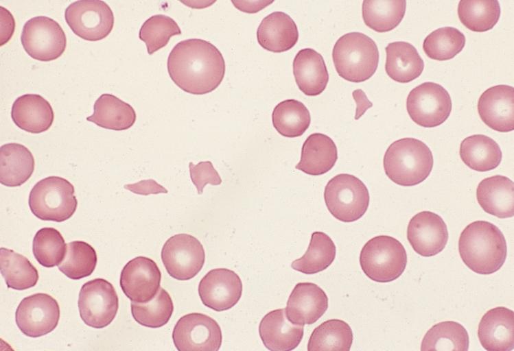
-RBC abnormalities suspicious for hemolysis on peripheral smear include the following:  spherocytes, schistocytes, helmet cells, acanthocytes, blister or bite cells, RBC with inclusions (as in malaria), teardrop RBC, Red Cell Ghosts,  and prominent basophilic stippling


-Iron Deficiency Anemia-


-presents as a microcytic hypochromic anemia
-with iron deficiency anemia, serum iron is reduced, the level of transferrin and TIBC is elevated

-25 percent of the worlds population is anemic and half of this population is due to iron deficiency anemia

-Most common in preschool children and women

-Reduced availability of iron is the most important cause of anemia due to impaired erythropoiesis

-There are two major causes of iron restricted erythropoiesis
1.  Absolute Iron Deficiency-iron stores in the bone marrow and other parts of the monocyte macrophage system in the spleen and liver are absent. Can result from poor intake of iron, reduced iron absorption, and increased blood loss
2.  Functional Iron Deficiency-with this there is insufficient availability of iron (low plasma iron levels and/or low transferrin saturation) for incorporation into erythroid precursors with normal or increased body iron stores.  This is the case of anemia of chronic disease (inflammation) or treatment with EPO for those patients with renal failure

-Causes of Iron Deficiency-
1.  Blood loss
2.  Decreased Iron Absorption (Celiac diseases or foods and medications blocking)
3.  Intravascular Hemolysis
4.  Pulmonary Hemosiderosis (Chronic Pulmonary Hemorrhage)
5.  Response to Erythropoietin
6.  Gastric Bypass for Morbid Obesity
7.  Congenital Iron Deficiency

-Clinical Symptoms include weakness, headache, irritability, and degrees of fatigue and exercise intolerance

-A presumptive diagnosis of iron deficiency anemia is made when there is a positive response of a trial of oral iron therapy.
-A positive response is a modest reticulocyte increase after 5-7 days and an increase in hemoglobin at a rate of approximately 2-4 g/dL every 3-4 weeks until hemoglobin is normal

-If there is no adequate response to iron trial may consider other diagnosis such as sprue, atrophic gastritis, gastrectomy, and from gastric bypass procedures.  May need IV iron treatments

-Other indications for parenteral iron include excessive continued blood loss, inflammatory bowel disease, chronic kidney disease, cancer patients, and heart failure patients.

-Bleeding Scan can assess for blood loss source

-Serum or plasma ferritin has replaced assessment of bone marrow stores for the diagnosis of absolute iron deficiency

-Need to consider malignancy or chronic kidney disease with microcytic hypochromic iron deficiency anemia


-Sickle Cell Anemia-


-Sickle Cell Disease is a vaso-occlusive phenomena and it causes hemolysis

-Sickle Cell Disease is an inherited disorder due to homozygous abnormal hemoglobin S (Hb S)

-Vasooclusion results in recurrent painful episodes (sickle cell crisis)

-The term sickle cell disease refers to all conditions associated with sickling, whereas sickle cell anemia describes the homozygous for hemoglobin S

-Patients with sickle beta thalassemia, the disease varies with the quantity of hemoglobin A, often being quite severe in patients with hemoglobin beta (0) thalassemia, and less severe in patients with sickle cell beta (+) thalassemia

-Among patients with coexisting alpha thalassemia, the anemia is less severe but the effects on clinical manifestations are variable

-Episodes of acute pain are the most common type of vasooclusive event and is the most common symptom after the age of 2.  Pain can be precipitated by weather conditions, dehydration, stress, menses, alcohol consumption, nocturnal hypoxemia, and rare obstructive sleep apnea.

-Half of vasooclusive have objective findings such as fever, swelling, tenderness, tachypnea, hypertension, nausea, and vomiting

-There is no test to diagnose a vasooclusive event.  The peripheral blood smear typically reveals that 5-50 percent of RBC that are reversible sickled cells

-Approximately 24 percent of individuals may suffer an overt stroke with sickle cell anemia by age of 45

-Multiorgan failure is most often seen during severe pain episodes in patients with sickle cell disease

-Reduced (hyposplenism) or absent splenic function is most commonly seen in subjects with sickle cell disease due to repeated infarction.  Children are susceptible to life threatening infections (Strep Pneumonia) and should be covered with rocephin parenterally

-Other complications of sickle cell disease include:  susceptibility to H. Influenza Type B, Meningitis, Bacterial Pneumonia, Osteomyelitis, bone infarction, and CVA

-Leg ulcers are a frequent complication of sickle cell disease

-Other complications include acute hepatic ischemia, benign cholestasis, hepatic sequestration crisis, transfusional iron overload, acute on chronic cholelithiasis with pigmented stones, drug toxicity, priapism, pulmonary complications, renal complications, retinopathy, and acute chest syndrome

-On lab evaluation will see mild to moderate anemia, increased reticulocytes, unconjugated hyperbilirubinemia, elevated LDH, and low serum haptoglobin.

-All individuals should receive appropriate vaccinations including these for strep pneumonia, influenza, neisseria meningitis, and HIB vaccine

-All individuals should receive folic acid supplementation.  Most symptomatic patients should be treated with hydroxyurea.

-During an acute pain episode patients should be treated with IV fluids, oxygen and opioid analgesics


-Thalassemia-


-Thalassemia refers to a spectrum of diseases characterized by reduced or absent production of one or more globin chains

-The majority of patients with alpha and beta thalassemia minor are asymptomatic and may be diagnosed because of microcytic hypochromic anemia

-Thalassemia of intermediate degrees of severity (thalassemia intermedia) are common throughout the world and may be due to the presence of more one hemoglobin mutation in the same patient or presence of an abnormal hemoglobin with a reduced production rate

-Beta thalassemia major and alpha thalassemia major are at the other end of the spectrum.  Beta thalassemia major are associated with lifelong transfusion dependent anemia.  Alpha is not compatible with extrauterine life

-Patients with Beta Thalassemia Major suffer from severe chronic anemia, chronic hemolysis, organ damage associated with iron overload, and profound local and systemic effects of a rapidly and relentless expanding mass of bone marrow progenitors

-Clinical manifestations of Beta Thalassemia Major include skeletal changes, hepatomegaly, Splenomegaly, Enlarged Kidneys (because of hematopoiesis), hypogonadism, growth failure, diabetes, and hypothyroidism

-Cardiac manifestations include heart failure and fatal arrhythmias are a manifestation of Beta Thalassemia Major

-Lab findings include profound hypochromic, microcytic anemia with bizarre morphology (Heinz Bodies)

-Iron level is usually elevated because of the high rate of RBC turnover, transferrin and TIBC is elevated

-Increased unconjugated bilirubin and LDH and low levels of haptoglobin are seen with Beta Thalassemia Major

-Diagnosis of Beta Thalassemia Major will be made in all patients around 6-12 months due to the presence of pallor, irritability, growth retardation, and abdominal swelling due to hepatomegaly and jaundice


-Vitamin B12 Deficiency-


-Vitamin B 12 Deficiency is dependent on dietary intake, stomach acid, and intrinsic factor in stomach to liberate Vitamin B12 from binding proteins, pancreatic proteases to free Vitamin B12 from binding R factors, secretion of intrinsic factor by gastric parietal cells to bind Vitamin B 12, and an intact ileum with functional receptors

-Causes of Vitamin B 12 Deficiency include pernicious anemia, autoimmune metaplastic atrophic gastritis, autoantibody formation against intrinsic factor, gastrectomy, gastritis, tapeworm, drugs, inadequate dietary intake, HIV infection, Nitrous oxide exposure,  and H. Pylori infection,  and hereditary problems

-Clinical manifestations include macrocytic anemia
-Only Vitamin B 12 deficiency produces neurologic changes not folate deficiency
-Neurologic changes include neuropathy, degeneration of the dorsal an lateral spine columns, weakness, spasticity, clonus, paraplegia, and even fecal and urinary incontinence can occur from deficiency

-Vitamin B 12 deficiency increases risk for osteoporosis

-Treatment involves depo preparation of Vitamin B 12 once a month and exploring possible correctable etiology